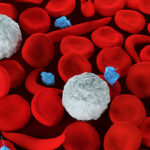
Wiskott-Aldrich syndrome (WAS) is a rare X-linked primary immunodeficiency that is characterized by eczema, recurrent infections, and micro-thrombocytopenia.
Wiskott-Aldrich syndrome is caused by an X-linked genetic defect in the WAS gene, which is located on short arm of the X-chromosome at Xp 11.22-23 position.
The gene product Wiskott-Aldrich protein (WASp) is a 502 amino acid protein that is expressed in the cytoplasm of non-erythroid hematopoietic cells.
More than 300 gene mutations have been identified that result in abnormal protein configuration.
Wiskott-Aldrich syndrome is caused by mutations in the gene WASP (WAS protein), which is involved in actin polymerization, cellular signaling, cytoskeletal rearrangement, and immunological synapse formation.
Individuals with the disease may experience a different number and severity of symptoms.
- Bleeding
- Immunodeficiency
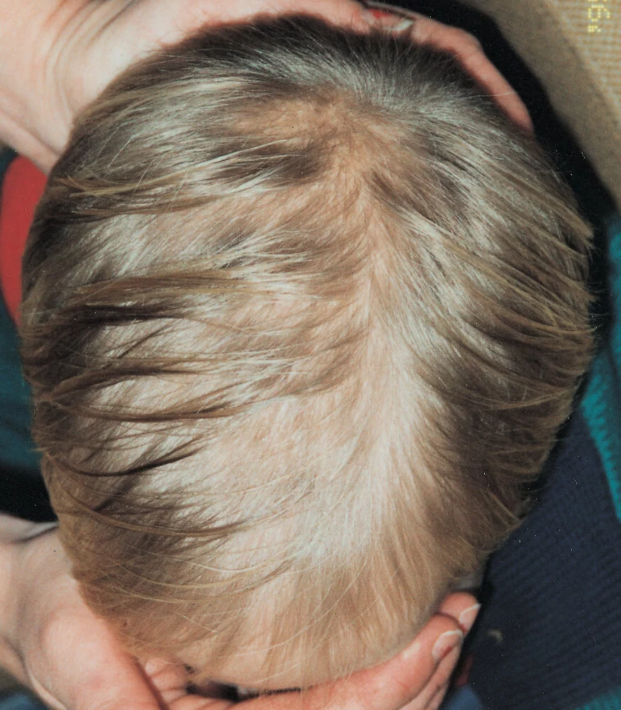
- Eczema
- Autoimmune Manifestations
- Malignancies
There are three main clinical phenotypic symptoms: –
- Classic (severe) Wiskott-Aldrich syndrome
- X-linked Neutropenia (XLN)
- X-linked Thrombocytopenia (XLT)
Any male with thrombocytopenia, eczema, recurrent respiratory infections, autoimmunity should be evaluated for WAS.
Any male patient with severe congenital neutropenia should be evaluated for XLN.
Abnormal immunologic findings in patients with WAS include: –
- Decrease number and function of T cells and regulatory T cells.
- Abnormal immunoglobulin (Ig) isotypes.
- Defective antigen-antibody response.
- Impaired cytotoxicity of natural killer cells with normal to increased cell numbers.
- Impaired chemotaxis of neutrophils and phagocytic cells.
- Absolute lymphocyte counts are typically normal during infancy, but T and B cell counts decline later in life in people with classic WAS.
- Variations in Ig levels, including normal serum IgG levels, decreasing IgM levels, and elevated IgA and IgE levels.
Wiskott-Aldrich syndrome is primarily managed with supportive care, which includes the following: –
- Broad-spectrum antibiotics for bacterial infections.
- Antivirals/antifungals for viral and fungal infections respectively.
- Platelet transfusions to prevent bleeding.
- Topical steroids to treat eczema.
Patients with significant antibody deficiency who have WAS or XLT should receive intravenous immunoglobin (IVIG) therapy.
Immunosuppressive therapy may be required for autoimmune manifestations.
In some patients, an elective splenectomy has been suggested as a way to stop the bleeding tendency and reverse the thrombocytopenia by increasing the number of circulating platelets.
HCT is the only available curative treatment.
Gene therapy is a potential alternative treatment for WAS that is currently being researched.